Jan 30, 2010

What is a Heart Attack?

Heart With Muscle Damage and a Blocked Artery

A heart attack occurs when blood flow to a section of heart muscle becomes blocked. If the flow of blood isn’t restored quickly, the section of heart muscle becomes damaged from lack of oxygen and begins to die.
Fortunately, today there are excellent treatments for heart attack that can save lives and prevent disabilities. Treatment is most effective when started within 1 hour of the beginning of symptoms.

Heart attacks occur most often as a result of a condition called coronary artery disease (CAD). In CAD, a fatty material called plaque builds up over many years on the inside walls of the coronary arteries (the arteries that supply blood and oxygen to your heart). Eventually, an area of plaque can rupture, causing a blood clot to form on the surface of the plaque. If the clot becomes large enough, it can mostly or completely block the flow of oxygen-rich blood to the part of the heart muscle fed by the artery.

Figure A is an overview of a heart and coronary artery showing damage (dead heart muscle) caused by a heart attack. Figure B is a cross-section of the coronary artery with plaque buildup and a blood clot.
During a heart attack, if the blockage in the coronary artery isn’t treated quickly, the heart muscle will begin to die and be replaced by scar tissue. This heart damage may not be obvious, or it may cause severe or long-lasting problems.
Severe problems linked to heart attack can include heart failure and life-threatening arrhythmias (irregular heartbeats). Heart failure is a condition in which the heart can’t pump enough blood throughout the body. Ventricular fibrillation is a serious arrhythmia that can cause death if not treated quickly.
Acting fast at the first sign of heart attack symptoms can save your life and limit damage to your heart. Treatment is most effective when started within 1 hour of the beginning of symptoms.
The most common heart attack signs and symptoms are:
  • Chest discomfort or pain—uncomfortable pressure, squeezing, fullness, or pain in the center of the chest that can be mild or strong. This discomfort or pain lasts more than a few minutes or goes away and comes back.

  • Upper body discomfort in one or both arms, the back, neck, jaw, or stomach.

  • Shortness of breath may occur with or before chest discomfort.

  • Other signs include nausea (feeling sick to your stomach), vomiting, lightheadedness or fainting, or breaking out in a cold sweat.

Jan 28, 2010

WHAT IS AIDS?


WHAT DOES "AIDS" MEAN?
AIDS stands for Acquired Immune Deficiency Syndrome:
Acquired means you can get infected with it;
Immune Deficiency means a weakness in the body's system that fights diseases.
Syndrome means a group of health problems that make up a disease.

AIDS is caused by a virus called HIV, the Human Immunodeficiency Virus. If you get infected with HIV, your body will try to fight the infection. It will make "antibodies," special molecules to fight HIV.

A blood test for HIV looks for these antibodies. If you have them in your blood, it means that you have HIV infection. People who have the HIV antibodies are called "HIV-Positive.

Being HIV-positive, or having HIV disease, is not the same as having AIDS. Many people are HIV-positive but don't get sick for many years. As HIV disease continues, it slowly wears down the immune system. Viruses, parasites, fungi and bacteria that usually don't cause any problems can make you very sick if your immune system is damaged. These are called "opportunistic infections."

HOW DO YOU GET AIDS?
You don't actually "get" AIDS. You might get infected with HIV, and later you might develop AIDS. You can get infected with HIV from anyone who's infected, even if they don't look sick and even if they haven't tested HIV-positive yet. The blood, vaginal fluid, semen, and breast milk of people infected with HIV has enough of the virus in it to infect other people. Most people get the HIV virus by:
* having sex with an infected person
* sharing a needle (shooting drugs) with someone who's infected
* being born when their mother is infected, or drinking the breast milk of an infected woman

Getting a transfusion of infected blood used to be a way people got AIDS, but now the blood supply is screened very carefully and the risk is extremely low.

There are no documented cases of HIV being transmitted by tears or saliva, but it is possible to be infected with HIV through oral sex or in rare cases through deep kissing, especially if you have open sores in your mouth or bleeding gums.

WHAT HAPPENS IF I'M HIV POSITIVE?
You might not know if you get infected by HIV. Some people get fever, headache, sore muscles and joints, stomach ache, swollen lymph glands, or a skin rash for one or two weeks. Most people think it's the flu. Some people have no symptoms.

The virus will multiply in your body for a few weeks or even months before your immune system responds. During this time, you won't test positive for HIV, but you can infect other people.

When your immune system responds, it starts to make antibodies. When this happens, you will test positive for HIV.

After the first flu-like symptoms, some people with HIV stay healthy for ten years or longer. But during this time, HIV is damaging your immune system.

One way to measure the damage to your immune system is to count your CD4 cells you have. These cells, also called "T-helper" cells, are an important part of the immune system. Healthy people have between 500 and 1,500 CD4 cells in a milliliter of blood.

Without treatment, your CD4 cell count will most likely go down. You might start having signs of HIV disease like fevers, night sweats, diarrhea, or swollen lymph nodes. If you have HIV disease, these problems will last more than a few days, and probably continue for several weeks.

HOW DO I KNOW IF I HAVE AIDS?
HIV disease becomes AIDS when your immune system is seriously damaged. If you have less than 200 CD4 cells or if your CD4 percentage is less than 14%, you have AIDS. See Fact Sheet 124 for more information on CD4 cells. If you get an opportunistic infection, you have AIDS. There is an "official" list of these opportunistic infections put out by the Centers for Disease Control (CDC). The most common ones are:
* PCP (Pneumocystis pneumonia), a lung infection;
* KS (Karposi's sarcoma), a skin cancer;
* CMV (Cytomegalovirus), an infection that usually affects the eyes
* Candida, a fungal infection that can cause thrush (a white film in your mouth) or infections in your throat or vagina

AIDS-related diseases also includes serious weight loss, brain tumors, and other health problems. Without treatment, these opportunistic infections can kill you.

AIDS is different in every infected person. Some people die a few months after getting infected, while others live fairly normal lives for many years, even after they "officially" have AIDS. A few HIV-positive people stay healthy for many years even without taking antiretroviral medications (ARVs).

IS THERE A CURE FOR AIDS?
There is no cure for AIDS. There are drugs that can slow down the HIV virus, and slow down the damage to your immune system. There is no way to "clear" the HIV out of your body.

Other drugs can prevent or treat opportunistic infections (OIs). In most cases, these drugs work very well. The newer, stronger ARVs have also helped reduce the rates of most OIs. A few OIs, however, are still very difficult to treat. See Fact Sheet 500 for more information on opportunistic infections.

Jan 27, 2010

Transpiration

Transpiration is the evaporation of water from plants. It occurs chiefly at the leaves while their stomata are open for the passage of CO2 and O2 during photosynthesis.

But air that is not fully saturated with water vapour (100% relative humidity) will dry the surfaces of cells with which it comes in contact. So the photosynthesizing leaf loses substantial amount of water by evaporation. This transpired water must be replaced by the transport of more water from the soil to the leaves through the xylem of the roots and stem.

Importance
Transpiration is not simply a hazard of plant life. It is the "engine" that pulls water up from the roots to:
- supply photosynthesis (1%-2% of the total)
- bring minerals from the roots for biosynthesis within the leaf
- cool the leaf
 
Environmental factors that affect the rate of transpiration

1. Light
Plants transpire more rapidly in the light than in the dark. This is largely because light stimulates the opening of the stomata (mechanism). Light also speeds up transpiration by warming the leaf.

2. Temperature
Plants transpire more rapidly at higher temperatures because water evaporates more rapidly as the temperature rises. At 30°C, a leaf may transpire three times as fast as it does at 20°C.

3. Humidity
The rate of diffusion of any substance increases as the difference in concentration of the substances in the two regions increases.When the surrounding air is dry, diffusion of water out of the leaf goes on more rapidly.

4. Wind
When there is no breeze, the air surrounding a leaf becomes increasingly humid thus reducing the rate of transpiration. When a breeze is present, the humid air is carried away and replaced by drier air.

5. Soil water
A plant cannot continue to transpire rapidly if its water loss is not made up by replacement from the soil. When absorption of water by the roots fails to keep up with the rate of transpiration, loss of turgor occurs, and the stomata close. This immediately reduces the rate of transpiration (as well as of photosynthesis). If the loss of turgor extends to the rest of the leaf and stem, the plant wilts.

The volume of water lost in transpiration can be very high. It has been estimated that over the growing season, one acre of corn plants may transpire 400,000 gallons of water. As liquid water, this would cover the field with a lake 15 inches deep. An acre of forest probably does even better.


Plant Tissue Systems

The tissues of a plant are organized into three tissue systems: the dermal tissue system, the ground tissue system, and the vascular tissue system.

Dermal Tissue System - Epidermis, Periderm (in older stems and roots)
• protection
• prevention of water loss

Ground Tissue System - Parenchyma tissue, Collenchyma tissue, Sclerenchyma tissue
• photosynthesis
• food storage
• regeneration
• support
• protection

Vascular Tissue System - Xylem, Phloem
• transport of water and minerals
• transport of food

Jan 23, 2010

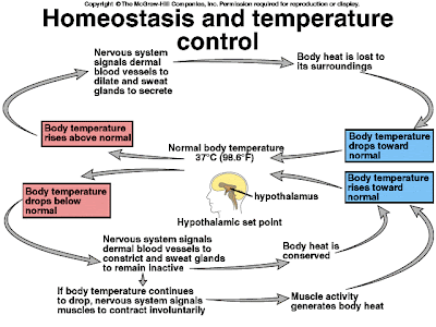
Homeostasis

Homeostasis is the maintenance of a relatively stable internal condition . The liver, the kidneys, and the brain (hypothalamus, the autonomic nervous system and the endocrine system) help maintain homeostasis. The liver is responsible for metabolising toxic substances and maintaining carbohydrate metabolism. The kidneys are responsible for regulating blood water levels, re-absorption of substances into the blood, maintenance of salt and ion levels in the blood, regulation of blood pH, and excretion of urea and other wastes.

An inability to maintain homeostasis may lead to death or a disease, a condition known as homeostatic imbalance. For instance, heart failure may occur when negative feedback mechanisms become overwhelmed and destructive positive feedback mechanisms take over. Other diseases which result from a homeostatic imbalance include diabetes, dehydration, hypoglycemia, hyperglycemia, gout and any disease caused by the presence of a toxin in the bloodstream.

Thermoregulation
Humans are warm-blooded, maintaining a near-constant body temperature. Thermoregulation is an important aspect of human homeostasis. Heat is mainly produced by the liver and muscle contractions. Humans have been able to adapt to a great diversity of climates, including hot humid and hot arid. High temperatures pose serious stresses for the human body, placing it in great danger of injury or even death. In order to deal with these climatic conditions, humans have developed physiologic and cultural modes of adaptation.

Temperature may enter a circle of positive feedback, when temperature reaches extremes of 45°C (113°F), at which cellular proteins denature, causing the active site in proteins to change, thus causing metabolism stop and ultimately death.

Glucoregulation


Humans regulate their blood glucose with insulin and glucagon. These hormones are released by the pancreas.

When blood sugar levels become too high, insulin is released from the pancreas, lowering the blood sugar levels. On the other hand, when blood sugar levels become too low, glucagon is released, increasing blood sugar levels.

If the pancreas is for any reason unable to produce enough of these two hormones, diabetes results.

Osmoregulation


Osmoregulation is the active regulation of the osmotic pressure of bodily fluids to maintain the homeostasis of the body's water content; that is it keeps the body's fluids from becoming too dilute or too concentrated. Osmotic pressure is a measure of the tendency of water to move into one solution from another by osmosis. The higher the osmotic pressure of a solution the more water wants to go into the solution.

The kidneys are used to remove excess ions from the blood, thus affecting the osmotic pressure. These are then expelled as urine

Volume
The body's homeostatic control mechanisms, which maintain a constant internal environment, ensure that a balance between fluid gain and fluid loss is maintained. The hormones ADH (Anti-diuretic Hormone, also known as vasopressin) and Aldosterone play a major role in this.

If the body is becoming fluid-deficient, there will be an increase in the secretion of these hormones (ADH), causing fluid to be retained by the kidneys and urine output to be reduced.

Conversely, if fluid levels are excessive, secretion of these hormones (aldosterone) is suppressed, resulting in less retention of fluid by the kidneys and a subsequent increase in the volume of urine produced.

If you have too much Carbon dioxide(CO2) in the blood, it can cause the blood to become acidic. People respirate heavily not due to low oxygen(O2) content in the blood, but because they have too much CO2.

Jan 22, 2010

Fluid Exchange Between Capillaries and Tissues


Capillaries are composed of a single layer of squamos epithelium surrounded by a thin basement membrane. Most capillaries (except those servicing the nervous system) have pores (spaces) between the individual cells that make up the capillary wall. Plasma fluid and small nutrient molecules leave the capillary and enter the interstitial fluid through these pores, in a process called bulk flow. Bulk flow facilitates the efficient transfer of nutrient out of the blood and into the tissues. However, blood cells and plasma proteins, which are too large to fit through the pores, do not filter out of the capillaries by bulk flow.

Together, blood plasma and interstitial fluid make up the extracellular fluid (ECF). Plasma constitutes 20%, while interstitial fluid constitutes 80% of the ECF. The distribution of extracellular fluid between these two compartments is determined by the balance between two opposing forces: hydrostatic pressure and osmotic pressure.


The beating of the heart generates hydrostatic pressure, which, in turn, causes bulk flow of fluid from plasma to interstitial fluid through walls of the capillaries. In other words, the pressure in the system forces plasma to filter out into the interstitial compartment. The composition of the interstitial fluid and the plasma is essentially the same except that plasma also contains plasma proteins not found in the interstitial fluid. Because of the presence of plasma proteins, the plasma has a higher solute concentration than does the interstitial fluid. Consequently, osmotic pressure causes interstitial fluid to be absorbed into the plasma compartment. In other words, the plasma proteins drive the reabsorption of water back into the capillaries via osmosis.

 
The magnitudes of filtration and absorption are not equal. The net filtration of fluid out of the capillaries into the interstitial compartment is greater than the net absorption of fluid back into the capillaries. The excess filtered fluid is returned to the blood stream via the lymphatic system. In addition to its roles in digestion and immunity, the lymphatic system functions to return filtered plasma back to the circulatory system. The smallest vessels of the lymphatic system are the lymphatic capillaries (shown in yellow). These porous, blind-ended ducts form a large network of vessels that infiltrate the capillary beds of most organs. Excess interstitial fluid enters the lymphatic capillaries to become lymph fluid.


Lymphatic capillaries converge to form lymph vessels that ultimately return lymph fluid back to the circulatory system via the subclavian vein. The presence of one-way valves in the lymph vessels ensures unidirectional flow of lymph fluid toward the subclavian vein.

If excess fluid cannot be returned to the blood stream then interstitial fluid builds up, leading to swelling of the tissues with fluid, this is called oedema.

Causes of Oedema

1. Reduced concentration of plasma proteins. When the concentration of plasma proteins drops, the osmotic potential of plasma drops, thus less interstitial fluid is absorbed into the capillaries. The rate of filtration, however, remain unchanged. Therefore, the ratio of filtration to absorption increases, leading to a build up of interstitial fluid. Any condition that would lead to a reduction in plasma proteins could potentially cause edema. Examples of conditions that reduce plasma proteins include:
     a) Kidney disease can result in the loss of plasma proteins in the urine.
     b) Liver disease can decrease the synthesis of plasma proteins.
     c) A protein-deficient diet will decrease plasma proteins.
     d) Severe burns result in a loss of plasma proteins (albumin) at the burn site

2. Increased capillary permeability. During an inflammatory response, tissue damage leads to the release of histamine from immune cells. Histamine causes an increase in the size of capillary pores. As capillaries become more permeable, the rate of filtration increases.

3. Increase in venous pressure. If venous pressure is increased then blood dams up in the upstream capillary bed, resulting in excess filtration. Examples of this condition include:

     a) Left heart failure. The left half of the heart drains blood from the lungs. When the left ventricle fails to adequately pump blood, venous pressure in the lungs increases. This increases in hydrostatic pressure causes an increase in the rate of filtration of fluid out of the capillaries and into the interstitial compartment. As a result, the lungs fill with fluid, a condition called, pulmonary oedema.

     b) Standing still. If one stands still for long period of time, then blood will pool in the veins of the legs. This will increase venous pressure and lead to weeping of fluid into the tissues. You can actually feel your feet swell if you stand motionless for a long time.

4. Blocked Lymphatic Vessels. If lymph vessels become blocked, then lymph fluid will not be drained from the affected area and the area will swell. Any condition that causes blockage or removal of lymph vessels can lead to oedema. Examples of this condition include:

     a) Filaria round worms are transmitted to humans by some species of mosquitoes. The worms migrate to the lymph vessels and block them. This causes dramatic swelling of the affected area, a condition called elephantiasis.

     b) Treatment for breast cancer may include removal of lymph vessels from breast and arms. This is done to limit the metastasis (spread) of cancerous cells to other parts of the body through the lymph. Removal of lymph vessels results in swelling of the affected area.

Jan 21, 2010

Time Management


"Perhaps the most valuable result of all education is the ability to make yourself do the things that you have to do, when it ought to be done, whether you like it or not." - Thomas Huxley

How many times have you planned to do something only to procrastinate or fail to carry out the plan? While there are many explanations for your failure to implement your plan, analysis of this failure has to start with the plan itself. Even if you do not want to do the work, to be successful it is necessary to stay motivated and make yourself do what needs to be done. Many students often say they will "try" to do the work, but unfortunately the definition of "try" in daily English means: "Forget it - I really don't want to do this and I won't do it. Further, when asked of my progress, I will employ excuses that relieve me of the responsibility to change the behaviour." All of these problems occur in school life. The good news is that you can employ effective time management strategies to overcome these obstacles and be successful throughout your career. These include planning, implementing the plan, and evaluating the plan.

Life in school has many distractions that can keep students from managing their time and keeping up with their studies. For example a boy named Nicky. Partying Nicky has tendencies to procrastinate and not manage his time wisely. In short, he is going to fail his subject if he does not shape up and plan his work and activities. Nicky needs to understand there are three main objectives that he must stick to or else his planning will fail. The first is to be ready for an exam 2-3 days before the test is administered. Second, use his daytime hours for studying, because he realises he wants to be with his friends in the evening. Lastly, he needs to distribute his studying hours so as not to cram. To plan his time successfully, he should compare his academic studies to a full-time job, thus leaving his night time hours for fun time. This will help him maintain a healthy balance between work and fun.

After Nicky plans his time he must implement the plan. To implement his plan he needs to stick to his daily and weekly schedule. One way to do this is to identify precise products he will produce during specified time periods. When he completes a task, he crosses it off the list. He should plan to study between classes, when the ideas are still fresh from the class. Further, he should visualise completing the task successfully. Visualisation, properly applied, is a powerful strategy. He should also keep in mind - two hours of studying during the day for every one hour of class time is far more beneficial than cramming for six hours the night before the exam.

Lastly, Nicky needs to monitor himself and his work. For example, he could pretend he is the employer at a large manufacturing corporation. According to his plans and how well he implemented them, he must decide if he should receive a reward as a productive employee, or if he would be fired for poor performance. Put another way, Nicky could monitor himself by answering the following questions each night:

"If he worked in a corporation and he was the President, would he hire himself the way he behaved today?"

"Am I proud of my performance today?"

Effective Time Management strategies lead to success; ineffective Time Management strategies lead to failure. It's that simple. Time Management strategies help students to stay motivated and on top of their deadlines for projects, help eliminate procrastination, and help to balance academic and fun time to maintain a healthy lifestyle in school.

Jan 17, 2010

Camner nak study Bio nie???


1. Slow down !!
The flow of a biology book is not like the flow of a novel. A novel can be read effortlessly, smoothly and rapidly, but biology books cannot. If you are reading a novel and are somewhat distracted, you can still get the idea of what it is about. When you are not concentrating on biology you will get very little out of it, and it will seem more difficult than it really is.

2. Every word counts.
Biology books are usually not repetitive, so there is little chance of picking something up from reading on. Writers of biology texts believe that extra words and repeats get in the way of clarity.

3. It is best to tackle each chapter at least three times.
The first time you should skim the chapter, noting topic sentences, words in bold print, all tables, diagrams and summary charts. This is best read before the class. The second reading should be in more detail, studying each area and not proceeding until each section is understood. Reread each section as many times as necessary until you understand its meaning. Mastery can take minutes or hours or days. The last major reading is for writing down terms and definitions and important concepts (see #5 below).

4. Talk to yourself as you read.
Explain what you have read aloud and make up your own examples to better understand what you have read. Rereading the material aloud, especially in your own words helps clarify the information. Hearing yourself makes a lot of difference.

5. Words and symbols of biology have specific meanings.
Each time you come to a new term or concept, cover up the text and see if you can express the idea aloud in your own words. Write down all the words you do not know. Emphasise words in bold type. Whenever possible write out the definitions in your own words. Strive for understanding the definitions so that you can easily state them in your own words; you are more likely to remember them that way. By saying it out loud and writing it, you are more like to recall it later, when needed.

6. Study all diagrams and charts.
They condense a lot of valuable information. Cover up and see if you can visualise them.

7. Write as you read.
• During your first reading write nothing in the text.
• Do not highlight ¬ it slows down reading and it is often used as an excuse for not concentrating.
• In a later reading, call attention to important words or phrases by underlining them (do not overdo this). Complete sentences or paragraphs should be bracketed and not underlined.
• Write summarising statements to yourself in the margin.
• Make notes to yourself right in the text.
• Note questions that you need to have clarified.

• DO NOT WORRY ABOUT THE RESALE VALUE OF THE TEXT.

8. Record all key points on a separate sheet.

9. If there are study questions at the end of the chapters, be sure you can answer them. They are good practice for the exam.

10. Make flash cards with terminology and concepts.

11. Keep testing yourself on a separate sheet of paper.

12. Without looking back, write out and say aloud the important points.

13. Create tasks for yourself as you read the text.
After reading an example and working it out for yourself, try to think of other examples that would fit the idea being discussed.

14. Use more than one book on the topic you are studying whenever possible.
Pick books that appeal to you. If you are very verbal, a book with long explanations is likely to be most helpful. If you are more visual, you might choose a book that has more illustrations.

15. Read the chapter before, and again after, class.
You will get the most out of class if you have read the material before the teacher presents it. Even if you feel that you understood the material in class, read it over again in the text. The more you review it the more likely you are to recall it.

16. If possible, have a friend or family member quiz you on your notes and text information.
Done regularly, this commits more information to long-term memory.

Good luck...........

Jan 16, 2010

Somethin to do with blood


The structure of blood vessels.


The composition of mammalian blood.


Differentiation of blood cells. Some of the pluripotent stem cells differentiate into lymphoid stem cells, which then develop into B cells and T cells, two types of lymphocytes that function in the immune response. All other blood cells differentiate from myeloid stem cells.


Blood clotting


Atherosclerosis. These light micrographs contrast a cross section of (a) a normal (healthy) artery with (b) an artery partially blocked by an atherosclerotic plaque. Plaques consist mostly of fibrous connective tissue and smooth muscle cells infiltrated with lipids.

Additional info

  • Arteriosclerosis is any hardening (and loss of elasticity) of large arteries and arterioles (small arteries). It is often due to hypertension.


  • Artherosclerosis is a hardening of an artery specifically due to an atheromatous plaque. Atherosclerosis is the most common form of arteriosclerosis. Atherosclerosis is characterised by a thickening of the intima with plaques that can contain lipid-laden macrophages ("foam cells"). The plaques contain free lipid (cholesterol, etc.) and are prone to calcification and ulceration.

Jan 14, 2010

Partial solar eclipse tomorrow!


Same as last year, the main show is not the partial eclipse, but the annular one which begins its path in Africa and passes through Chad, Central African Republic, Democratic Republic of the Congo, Uganda, Kenya, and Somalia. After leaving Africa, the path crosses the Indian Ocean where the maximum duration of annularity reaches 11 min 08 s. The central path then continues into Asia through Bangladesh, India, Myanmar, and China.

A partial eclipse is seen within the much broader path of the Moon’s penumbral shadow, which includes Eastern Europe, most of Africa, Asia, and Indonesia.

In Malaysia, the first contact – when the Moon first “touches” the Sun – begins around 3:01 pm. The Moon will then slowly cover up part of the Sun until maximum eclipse at 4:26 pm, when the Sun is 39 degrees above the horizon. Around 5:38 pm, the Moon leaves the Sun’s disk and the show ends.

During maximum eclipse, 28.6% of the Sun’s disk (area) will be obscured by the Moon, although 40.9% of the Sun’s diameter is obscured. That’s the different between eclipse obscuration and eclipse magnitude.

So now, how to observe this event safely?

There are few ways. You can project the image of the Sun onto a piece of paper or a wall either by using pinhole projector or telescope, or use a solar filter either the glasses type or attached it in front of a telescope. Only then, you can observe the Sun safely. You don’t really need a telescope to enjoy this.

Please bear in mind that although the Sun will be partially covered, the remaining of the crescent Sun will still be intense enough to damage your eyes. It is NOT safe at all to look at the partial Sun directly.

Jan 12, 2010

You Can Be Whatever You Want To Be


There is inside you all of the potential to be

whatever you want to be...

all of the energy to do whatever you want to do.

Imagine yourself as you would like to be,

doing what you want to do,

and each day, take one step towards your dream.

And though at times it may seem too difficult to continue,

hold on to your dream.

One morning you will awake to find

that you are the person you dreamed of...

doing what you wanted to do...

simply because you had the courage

to believe in your potential

and to hold on to your dream.

~Donna Levine~

Davydenko pips Nadal to win Qatar Open


Nikolay Davydenko saved two match points as he made a remarkable recovery to beat Rafael Nadal 0-6 7-6 6-4 in the Qatar Open final..

After failing to win a game in the opening set, and trailing in both the second and third, the Russian fought back strongly to overcome the world number two in one hour 43 minutes.
Third seed Davydenko, who defeated the world's top player Roger Federer in the semi-final, produced 52 winners against the second-seeded Spaniard's 37 to claim the trophy.
The world number six thought he had no chance after losing the first set without winning a game but a battling performance improved his head-to-head record against Nadal to 5-4.
"After losing the first set 6-0, I thought I had no chance of winning," said the Russian. "In the beginning, he was on top and I had to fight for every point. However, as the match wore on, Rafa lost his concentration and made too many mistakes.
"Really, I wasn't sure of winning till I had three match points. If I can make such a remarkable turnaround after failing completely in the first set that definitely means my confidence is high. I'm happy I could stay focussed at crucial stages."

UNBEATEN RUN
The victory takes Davydenko's unbeaten run to nine matches after he won the ATP World Tour Finals in London in November.
Nadal tried to look for positives as he prepares for the defence of his Australian Open title later this month.
"I'm happy with the way I performed. I played unbelievable tennis in the first set. That just shows I'm back to my best, maybe, like how I played in 2008. It's a huge morale booster," he said.
"After losing the tiebreaker (10-8), I don't know what went wrong. I lost to one of the best players in the world," Nadal said, adding it was too early to comment on his title prospects at the Australian Open.
Davydenko said beating Nadal and Federer at the Australian Open would be a completely different challenge.

Jan 11, 2010

Circulation


In insects, other arthropods, and most molluscs, blood bathes the organs directly in an open circulatory system.
There is no distinction between blood and interstitial fluid, and this general body fluid is more correctly termed hemolymph. One or more hearts pump the hemolymph into an interconnected system of sinuses, which are spaces surrounding the organs. Here, chemical exchange occurs between the hemolymph and body cells. In insects and other arthropods, the heart is an elongated tube located dorsally. When the heart contracts, it pumps hemolymph through vessels out into sinuses. When the heart relaxes, it draws hemolymph into the circulatory system through pores called ostia. Body movements that squeeze the sinuses help circulate the hemolymph.


The mammalian cardiovascular system : Note that the dual circuits operate simultaneously, not in the serial fashion that the numbering in the diagram suggests. The two ventricles pump almost in unison; while some blood is traveling in the pulmonary circuit, the rest of the blood is flowing in the systemic circuit.


Located beneath the breastbone (sternum), the human heart is about the size of a clenched fist and consists mostly of cardiac muscle. The two atria have relatively thin walls and serve as collection chambers for blood returning to the heart, most of which flows into the ventricles as they relax. Contraction of the atria completes filling of the ventricles. The ventricles have thicker walls and contract much more strongly than the atria—especially the left ventricle, which must pump blood to all body organs through the systemic circuit.


The cardiac cycle. For an adult human at rest with a pulse of about 75 beats per minute, one complete cardiac cycle takes about 0.8 second. 1 During a relaxation phase (atria and ventricles in diastole), blood returning from the large veins flows into the atria and ventricles. 2 A brief period of atrial systole then forces all remaining blood out of the atria into the ventricles. 3 During the remainder of the cycle, ventricular systole pumps blood into the large arteries. Note that seven–eighths of the time—all but 0.1 second of the cardiac cycle—the atria are relaxed and are filling with blood returning via the veins.


 A region of the heart called the sinoatrial (SA) node, or pacemaker, sets the rate and timing at which all cardiac muscle cells contract. Composed of specialized muscle tissue, the SA node is located in the wall of the right atrium, near the point where the superior vena cava enters the heart. Because the pacemaker of the human heart (and of other vertebrates) is made up of specialized muscle tissues and located within the heart itself, the vertebrate heart is referred to as a myogenic heart.
The SA node generates electrical impulses much like those produced by nerve cells. Because cardiac muscle cells are electrically coupled (by the intercalated disks between adjacent cells), impulses from the SA node spread rapidly through the walls of the atria, causing both atria to contract in unison.
The impulses also pass to another region of specialized cardiac muscle tissue, a relay point called the atrioventricular (AV) node, located in the wall between the right atrium and right ventricle. Here the impulses are delayed for about 0.1 second before spreading to the walls of the ventricles. The delay ensures that the atria empty completely before the ventricles contract. Specialized muscle fibers called bundle branches and Purkinje fibers then conduct the signals to the apex of the heart and throughout the ventricular walls.

The impulses that travel through cardiac muscle during the heart cycle produce electrical currents that are conducted through body fluids to the skin, where the currents can be detected by electrodes and recorded as an electrocardiogram (ECG or EKG).

Jan 9, 2010

Animal and Plant Cells


TSA/V Ratio


Geometric relationships between surface area and volume. In this diagram, cells are represented as boxes. Using arbitrary units of length, we can calculate the cell’s surface area (in square units), volume (in cubic units), and ratio of surface area to volume. The smaller the cell, the higher the surface–to–volume ratio. A high surface–to–volume ratio facilitates the exchange of materials between a cell and its environment.

Plant Tissues


A mature vascular plant, e.g., a tobacco plant, contains several differentiated cell types. These are grouped together in tissues. Some tissues contain only one type of cell. Some consist of several.


Meristematic

The main function of meristematic tissue is mitosis. The cells are small, thin-walled, with no central vacuole and no specialised features.

Meristematic tissue is located in
-the apical meristems at the growing points of roots and stems.
-the secondary meristems (lateral buds) at the nodes of stems (where branching occurs), and in some plants,
-a ring of meristematic tissue, called the cambium, that is found within the mature stem.

The cells produced in the meristems soon become differentiated into one or another of several types.

Protective

Protective tissue covers the surface of leaves and the living cells of roots and stems. Its cells are flattened with their top and bottom surfaces parallel. The upper and lower epidermis of the leaf are examples of protective tissue.

Parenchyma

The cells of parenchyma are large, thin-walled, and usually have a large central vacuole. They are often partially separated from each other. They are usually stuffed with plastids.

In areas not exposed to light, colorless plastids predominate and food storage is the main function. The cells of the white potato are parenchyma cells.

Where light is present, e.g., in leaves, chloroplasts predominate and photosynthesis is the main function.

Sclerenchyma

The walls of these cells are very thick and built up in a uniform layer around the entire margin of the cell. Often, the protoplasts die after the cell wall is fully formed. Sclerenchyma cells are usually found associated with other cells types and give them mechanical support.

Sclerenchyma is found in stems and also in leaf veins. Sclerenchyma also makes up the hard outer covering of seeds and nuts.

Collenchyma

Collenchyma cells have thick walls that are especially thick at their corners. These cells provide mechanical support for the plant. They are most often found in areas that are growing rapidly and need to be strengthened. The petiole ("stalk") of leaves is usually reinforced with collenchyma.

Xylem

Xylem conducts water and dissolved minerals from the roots to all the other parts of the plant. Link to discussion of water and mineral transport in the xylem.

In angiosperms, most of the water travels in the xylem vessels. These are thick-walled tubes that can extend vertically through several feet of xylem tissue. Their diameter may be as large as 0.7 mm. Their walls are thickened with secondary deposits of cellulose and are usually further strengthened by impregnation with lignin. The secondary walls of the xylem vessels are deposited in spirals and rings and are usually perforated by pits.

Xylem vessels arise from individual cylindrical cells oriented end to end. At maturity the end walls of these cells dissolve away and the cytoplasmic contents die. The result is the xylem vessel, a continuous nonliving duct. The vessels carry water and some dissolved solutes, such as inorganic ions, up the plant.

Xylem also contains tracheids. These are individual cells tapered at each end so the tapered end of one cell overlaps that of the adjacent cell. Like xylem vessels, they have thick, lignified walls and, at maturity, no cytoplasm. Their walls are perforated so that water can flow from one tracheid to the next. The xylem of ferns and conifers contains only tracheids.

In woody plants, the older xylem ceases to participate in water transport and simply serves to give strength to the trunk. Wood is xylem. When counting the annual rings of a tree, one is counting rings of xylem.

Phloem

The main components of phloem are
-sieve elements and
-companion cells.

Sieve elements are so-named because their end walls are perforated. This allows cytoplasmic connections between vertically-stacked cells. The result is a sieve tube that conducts the products of photosynthesis - sugars and amino acids - from the place where they are manufactured (a "source"), e.g., leaves, to the places ("sinks") where they are consumed or stored; such as

-roots
-growing tips of stems and leaves
-flowers
-fruits, tubers, corms, etc.

Sieve elements have no nucleus and only a sparse collection of other organelles. They depend on the adjacent companion cells for many functions.

Companion cells move sugars and amino acids into and out of the sieve elements. In "source" tissue, such as a leaf, the companion cells use transmembrane proteins to take up - by active transport - sugars and amino acids from the cells manufacturing them. Water follows by osmosis. These materials then move into adjacent sieve elements by diffusion through plasmodesmata. The pressure created by osmosis drives the flow of materials through the sieve tubes.

In "sink" tissue, the sugars and amino acids leave the sieve tubes by diffusion through plasmodesmata connecting the sieve elements to the cells of their destination. Again, water follows by osmosis where it may

-leave the plant by transpiration or
-increase the volume of the cells or
-move into the xylem for recycling through the plant.


Jan 4, 2010

World's tallest skyscraper


Blazing fireworks and dazzling lights marked the inauguration of the world's tallest tower, Burj Dubai.

The needle-shaped concrete, steel and glass tower, described by its developer as a "vertical city" as it dwarfs existing skyscrapers, boasts new limits in design and construction.

Emaar Properties, the partly government-owned developer, has maintained the suspense over the final height of the skyscraper, saying only that it exceeds 800 metres (2,625 feet).

But it revealed on that the tower will have over 200 floors, only 160 of which will be inhabited, while the remaining floors will be for services.

Burj Dubai has a total built-up area of 5.67 million square feet, including 1.85 million square feet of residential space and over 300,000 square feet of prime office space, Emaar said. Related article: History of Dubai mega projects

This amounts to 1,044 apartments and 49 floors of office space, served by 57 lifts.

A hotel carrying the Georgio Armani logo will also occupy part of the tower.

Bill Baker, a structural and civil engineer and partner in Chicago-based Skidmore, Owings and Merrill (SOM), which designed the tower, said Burj Dubai has set a new benchmark.

"We thought that it would be slightly taller than the existing tallest tower of Taipei 101. (Emaar) kept on asking us to go higher but we didn't know how high we could go," he said.

"We were able to tune the building like we tune a music instrument. As we went higher and higher and higher, we discovered that by doing that process... we were able to reach heights much higher than we ever thought we could.

"We learned quite a bit from Burj Dubai. I would think we could easily do a one kilometre (tower). We are optimistic about the ability to go even higher."

A spiralling Y-shaped design by SOM architect Adrian Smith was used to support the structural core of the tower, which narrows as it ascends. Higher up it becomes a steel structure topped with a huge spire. Related article: The world's highest towers.

To reach the final stages, concrete was propelled to a height of 605 metres (1,996 feet) -- a world record.

Jan 3, 2010

Exam Format for STPM Biology

Candidates are required to enter for Papers 1, 2, and either Paper 3 or Paper 4.

Paper 1
- 50 compulsory multiple-choice questions are to be answered.
- 50 marks (to be scaled to 60)
- 1¾ hours

Paper 2
Section A:
- 4 compulsory short structured questions are to be answered.
- 10 marks per question (40 marks)
Section B:
- 4 questions are to be answered out of 6 essay questions.
- 15 marks per question (60 marks)
- Total: 100 (to be scaled to 120)
- 2½ hours

Paper 3
School-based Assessment of Practical:
- 13 compulsory experiments are to be carried out.
- (20 marks)
- During school term

Practical Syllabus
School-based Assessment of Practical (Paper 3)

School-based assessment of practical work will only be carried out during the school term of form six for candidates from government and private schools which have been approved by the Malaysian Examinations Council to carry out the school-based assessment. Individual private candidates, candidates from private schools which have no permission to carry out the school-based assessment of practical work, candidates who repeat upper six (in government or private schools), and candidates who do not attend classes of lower six and upper six for two consecutive years (in government or private schools) are not allowed to take this paper.

13 compulsory experiments (including three projects) are to be carried out by candidates and to be assessed by subject teachers in schools. Candidates are required to carry out the projects individually or in groups as stipulated. Details of the topic, aim, theory, apparatus, and method of each of the experiments are with the teachers.

Students will be supplied with a work scheme before the day of the compulsory experiment so as to enable them to plan their practical work. Each experiment is expected to last one school double period. Assessment of the students’ practical work will be done by the teacher during the practical session and will also be based on the students’ practical report. The assessment should comply with the assessment guidelines prepared by the Malaysian Examinations Council.
 
Reference Books

1. Audesirk, T., Audesirk, G. & Bayers, B.E., Biology: Life on Earth, (6th ed.), Prentice-Hall, 2002.

2. Campbell, N. A. & Reece, J. B., Biology, (6th ed.), Benjamin Cummings, 2002.

3. Clegg, C. J. & Mackean, D. G., Advanced Biology: Principles and Applications, John Murray, 2000.

4. Green, N. P. O., Stout, G. W., & Taylor, D. J., Biological Science 1 & 2 (2nd ed.), Cambridge University Press, 1990.

5. Jones, M. & Jones, G., Advanced Biology, Cambridge University Press, 1997.

6. Solomon, E., P., Berg, L. R., & Martin, D. W., Biology, (6th ed.), Thomson Learning, 2002.

7. Starr C. & Taggart R., Biology: The Unity and Diversity of Life, (9th ed.), Von Hoffmen Press, 2000.

Exam Format for SPM Biology

Paper 1 (4551/1)
- Objective questions
- Multiple choice
- Each item consists of four choices of answers; A, B, C and D
- 50 questions (answer all)
- Total marks : 50
- Duration : 1 hour 15 mins
- Coverage of context : all area of learning

Paper 2 (4551/2)
- Section A : Subjective questions
   Section B : Essay writing
- Subjective : 5 items (answer all) - 60 marks
  Essay : 4 items (choose 2) - 40 marks
- Total marks : 100
- Duration : 2 hours 30 mins
- Coverage of context : all area of learning

Paper 3 (4551/3)
- Q1 : Structured - based item (33 marks)
  Q2 : Open response item (17 marks)
- Total marks : 50
- Duration : 1 hour 30 mins
- Coverage of context : Experiments

There you are, the SPM Biology Paper.
I'll share with you all next time the techniques in answering (especially Paper 2 and 3).
Most of the students are not familiar with the techniques and end up getting average marks.

Jan 2, 2010

What is Biology?


What is biology? Simply put, it is the study of life -- life in all of its grandeur. From the very small algae to the very large elephant, life has a certain wonder about it. With that in mind, how do we know if something is living? Is a virus alive or dead? What are the characteristics of life? These are all very important questions with equally important answers.

Characteristics of Life

Living things include both the visible world of animals and plants, as well as the invisible world of bacteria. On a basic level, we can say that life is ordered. Organisms have an enormously complex organization. We're all familiar with the intricate systems of the basic unit of life, the cell.

Life can also "work." No, not the daily employment variety, but living creatures can take in energy from the environment. This energy, in the form of food, is transformed to maintain metabolic processes and for survival.

Life grows and develops. This means more than just getting larger in size. Living organisms also have the ability to rebuild and repair themselves when injured.

Life can reproduce. Have you ever seen dirt reproduce? I don't think so. Life can only come from other living creatures.

Life can respond. Think about the last time you accidentally stubbed your toe. Almost instantly, you flinched back in pain. Life is characterized by this response to stimuli.

Finally, life can adapt and respond to the demands placed on it by the environment. There are three basic types of adaptations that can occur in higher organisms.

Reversible changes occur as a response to changes in the environment. Let's say you live near sea level and you travel to a mountainous area. You may begin to experience difficulty breathing and an increase in heart rate as a result of the change in altitude. These symptoms go away when you go back down to sea level.

Somatic changes occur as a result of prolonged changes in the environment. Using the previous example, if you were to stay in the mountainous area for a long time, you would notice that your heart rate would begin to slow down and you would begin to breath normally. Somatic changes are also reversible.

The final type of adaptation is called genotypic (caused by mutation). These changes take place within the genetic makeup of the organism and are not reversible. An example would be the development of resistance to pesticides by insects and spiders.

In summary, life is organized, "works," grows, reproduces, responds to stimuli and adapts. These characteristics form the basis of the study of biology.

Basic Principles of Biology

The foundation of biology as it exists today is based on five basic principles. They are the cell theory, gene theory, evolution, homeostasis, and laws of thermodynamics.

Cell Theory: all living organisms are composed of cells. The cell is the basic unit of life.

Gene Theory: traits are inherited through gene transmission. Genes are located on chromosomes and consist of DNA.

Evolution: any genetic change in a population that is inherited over several generations. These changes may be small or large, noticeable or not so noticeable.

Homeostasis: ability to maintain a constant internal environment in response to environmental changes.

Thermodynamics: energy is constant and energy transformation is not completely efficient.

Subdiciplines of Biology

The field of biology is very broad in scope and can be divided into several disciplines. In the most general sense, these disciplines are categorized based on the type of organism studied. For example, zoology deals with animal studies, botany deals with plant studies, and microbiology is the study of microorganisms. These fields of study can be broken down further into several specialized sub-disciplines. Some of which include anatomy, cell biology, genetics, and physiology.

The Lymphatic System


Closely connected with the blood and circulatory system, the lymphatic system is an extensive drainage system that returns water and proteins from various tissues back to the bloodstream. It is comprised of a network of ducts, called lymph vessels and carries lymph, a clear, watery fluid that resembles the plasma of blood. Some scientists consider this system to be part of the blood and circulatory system because lymph comes from blood and returns to blood, and because its vessels are very similar to the veins and capillaries of the blood system. Throughout the body, wherever there are blood vessels, there are lymph vessels, and the two systems work together.

How Are the Spleen and Lymphatic System Necessary for Living?

The entire lymphatic system flows toward the bloodstream, returning fluid from body tissues to the blood. If there were no way for excess fluid to return to the blood, our body tissues would become swollen. For example, when a body part swells, it may be because there is too much fluid in the tissues in that area. The lymph vessels collect that excess fluid and carry it to the veins through the lymphatic system.

This process is crucial because water, proteins, and other molecules continuously leak out of tiny blood capillaries into the surrounding body tissues. This lymph fluid has to be drained, and so it returns to the blood via the lymphatic vessels. These vessels also prevent the back flow of lymph fluid into the tissues.

The lymphatic system also helps defend the body against invasion by disease-causing agents such as viruses, bacteria, or fungi. Harmful foreign materials are filtered out by small masses of tissue called lymph nodes that lie along the network of lymphatic vessels. These nodes house lymphocytes (white blood cells), some of which produce antibodies, special proteins that fight off infection. They also stop infections from spreading through the body by trapping disease-causing germs and destroying them.

The spleen also plays an important part in a person's immune system and helps the body fight infection. Like the lymph nodes, the spleen contains antibody-producing lymphocytes. These antibodies weaken or kill bacteria, viruses, and other organisms that cause infection. Also, if the blood passing through the spleen carries damaged cells, white blood cells called macrophages in the spleen will destroy them and clear them from the bloodstream.

Basic Anatomy

The lymphatic system is a network of very fine vessels or tubes called lymphatics that drain lymph from all over the body. Lymph is composed of water, protein molecules, salts, glucose, urea, lymphocytes, and other substances.

Lymphatics are found in every part of the body except the central nervous system. The major parts of the system are the bone marrow, spleen, thymus gland, lymph nodes, and the tonsils. Other organs, including the heart, lungs, intestines, liver, and skin also contain lymphatic tissue.

Lymph nodes are round or kidney-shaped, and range in size from very tiny to 1 inch in diameter. They are usually found in groups in different places throughout the body, including the neck, armpit, chest, abdomen, pelvis, and groin. About two thirds of all lymph nodes and lymphatic tissue are within or near the gastrointestinal tract.

Lymphocytes are white blood cells in the lymph nodes that help the body fight infection by producing antibodies that destroy foreign matter such as bacteria or viruses. Two types are T-cells and B-cells. Some lymphocytes become stimulated and enlarged when they encounter foreign substances; these are called immunoblasts.

The major lymphatic vessel is the thoracic duct, which begins near the lower part of the spine and collects lymph from the lower limbs, pelvis, abdomen, and lower chest. It runs up through the chest and empties into the blood through a large vein near the left side of the neck. The right lymphatic duct collects lymph from the right side of the neck, chest, and arm, and empties into a large vein near the right side of the neck.

The spleen is found on the left side of the abdomen. Unlike other lymphoid tissue, red blood cells flow through it. It helps control the amount of blood and blood cells that circulate through the body and helps destroy damaged cells.

Normal Physiology

Lymph drains into open-ended, one-way lymph capillaries. It moves more slowly than blood, pushed along mainly by a person's breathing and contractions of the skeletal muscles. The walls of blood capillaries are very thin, and they have many tiny openings to allow gases, water, and chemicals to pass through to nourish cells and to take away waste products. Interstitial fluid passes out of these openings to bathe the body tissues.

Lymph vessels recycle the interstitial fluid and return it to the bloodstream in the circulatory system. They collect the fluid and carry it from all of the body's tissues and then empty it into large veins in the upper chest, near the neck.

Lymph nodes are made of a mesh like network of tissue. Lymph enters the lymph node and works its way through passages called sinuses. The nodes contain macrophages, phagocytic cells that engulf (phagocytize) and destroy bacteria, dead tissue, and other foreign matter, removing them from the bloodstream. After these substances have been filtered out, the lymph then leaves the nodes and returns to the veins, where it reenters the bloodstream.

When a person has an infection, germs collect in great numbers in the lymph nodes. If the throat is infected, for example, the lymph nodes of the neck may swell. Sometimes the phagocytic cells may not be able to destroy all of the germs, and a local infection in the nodes may result.

Because the lymphatic system extends to the far reaches of the body, it also plays a role in the spread of cancer. This is why lymph nodes near a cancerous growth are usually removed with the growth.

Diseases, Conditions, Disorders, and Dysfunction's

Because the lymphatic system branches through most of the parts of the body, it may be involved in a wide range of conditions. Diseases may affect the lymph nodes, the spleen, or the collections of lymphoid tissue that occur in certain areas of the body.

Disorders of the lymph nodes

Lymphadenopathy. Most lymph nodes in the body can't be felt easily unless they become swollen or enlarged. Lymphadenopathy is an increase in the size of a lymph node or nodes, most often as the result of a nearby infection (for example, lymphadenopathy in the neck might be the result of an infection of the throat). Less commonly (particularly in children), swelling of the lymph nodes can be due to an infiltration of cancerous cells. If lymphadenopathy is generalized (meaning that the swelling is present in several lymph node groups throughout the body), it usually indicates that the person has a systemic disease.

Lymphadenitis, or adenitis, is an inflammation (swelling, tenderness, and sometimes redness and warmth of the overlying skin) of the lymph node due to an infection of the tissue in the node itself. In children, this condition most commonly involves the lymph nodes of the neck.

Lymphomas. A group of cancers that arise from the lymph nodes, these diseases result when lymphocytes undergo changes and start to multiply out of control. The involved lymph nodes enlarge, and the cancer cells crowd out healthy cells and may form tumors (solid growths) in other parts of the body.

Disorders of the spleen

Splenomegaly (enlarged spleen). In children, the spleen is usually small enough that it can't be felt by pressing on the abdomen, but the spleen can enlarge to several times its normal size with certain diseases. There are many possible reasons for this including various blood diseases and cancers, but the most common cause in children is infection (particularly viral infections). Infectious mononucleosis, a condition usually caused by the Epstein-Barr virus (EBV), is one of many viral infections associated with an enlarged spleen. Children and teens with an enlarged spleen should avoid contact sports because they can have a life-threatening loss of blood if their spleen is ruptured.

Disorders of other lymphoid tissue

Tonsillitis. An extremely common condition, particularly in children, tonsillitis occurs when the tonsils, the collections of lymphoid tissue in the back of the mouth at the top of the throat, are involved in a bacterial or viral infection that causes them to become swollen and inflamed. The tonsils normally help to filter out bacteria and other microorganisms to aid the body in fighting infection. Symptoms include sore throat, high fever, and difficulty swallowing. The infection may also spread to the throat and surrounding areas, causing pain and inflammation (pharyngitis).

Glossary

antibodies:
chemicals produced by white blood cells to fight bacteria, viruses, and other foreign substances

immunoblasts:
Lymphocytes that become stimulated and enlarged when they encounter foreign substances

interstitial fluid:
fluid that leaks out of capillaries (the tiniest blood vessels) and bathes body tissues

lymph vessels:
channels or ducts that contain and convey lymph; also called lymphatics

lymph:
pale fluid that bathes the body tissues, passes into lymphatic vessels, and is discharged into the blood by way of the thoracic duct; it consists of a liquid resembling blood plasma and contains white blood cells

lymph nodes:
organized masses of lymphoid tissue that are distributed along the branching system of lymphatic vessels; they contain numerous lymphocytes and other cells that filter bacteria, dead tissue, and foreign matter from the lymph that flows through them

lymphocytes:
white blood cells

macrophages:
white blood cells that remove damaged cells from the bloodstream

spleen:
organ found on the left side of the abdomen; it helps control the amount of blood and blood cells that circulate through the body and helps destroy damaged cells

thoracic duct:
major lymphatic vessel, which begins near the lower part of the spine and collects lymph from the lower limbs, pelvis, abdomen, and lower chest; lymph flowing through the duct eventually empties into a large vein in the upper chest and returns to the bloodstream.